My Research
Rife Reference Page
Back To Top Of Page
Gaston Naessens' Microscope
What Rife accomplished optically in the 1930s with his Universal Microscope, Gaston Naessens accomplished with a combination of optics and electronics in the 1940s in his Somatoscope. Born on 16 March 1924 in Roubaix, France, Gaston displayed a predisposition to be an inventor when at the age of five he built a little moving auto-like toy from a Meccano set and powered it with an alarm clock spring. Later, he built a home-made motorcycle and a mini-airplane! While attending the University of Lille, Gaston nearly had his education disrupted by the German invasion. Fortunately, Gaston and his fellow students escaped to Nice where they carried on their education in exile. He was awarded a diploma from the Union Nationale Scientifique Francaise - a quasi-official institution under whose auspices the education of the displaced students continued. He did not bother seeking an equivalency degree from the de Gaulle government when French rule was restored. At the young age of twenty-one, frustrated by the limitations of conventional microscopes, Gaston set out to build a superior microscope. Technical assistance was provided by German craftsmen from Wetzlar, Germany, who checked out many of Gaston's ideas on optics. Privately, Gaston devised the electrical manipulation of the light source. Once the technical aspects were resolved, Gaston had the body of his microscope constructed by Barbier-Bernard et Turenne, technical specialists and defence contractors near Paris.
THEORY OF OPERATION: The Somatoscope mixes light from two orthogonal light sources - a mercury lamp and a halogen lamp. The light from both sources enters a glass tube at 90 degrees from each other. As the light waves beat against each other, a strong carrier wave of light emerges and travels down the light tube. (It should be noted that two electromagnetic fields superimposed on each other at 90 degrees is a classic scalar formation!) As the light travels down the tube, it passes through a monochromatic filter which forms it into a monochromatic ray. The ray is then passed through a large coil that surrounds the tube. The coil's magnetic field divides the ray into numerous parallel rays that are then passed through a Kerr cell which increases the frequency of the ray before being injected onto the specimen. The light, which contains the carrier and a mixture of selected signals in the UV range, stimulates the biological material in the Somatoscope to the point that the specimens give off their own light. (Rife referred to this as luminescence.) This is the key to the ultra-high resolution that has been achieved by Gaston Naessens. Conventional microscopes pass light through the specimen which theoretically limits the resolution of optical microscopes to the wavelength of light. The finest optical microscopes have achieved magnification levels of 2,500 diameters. At levels above this, the resolution is limited by the wavelength of light and further magnification merely creates a blur! Higher resolutions have been achieved by microscopes that do not use lenses, but instead use apertures which are smaller than the wavelength of light. One such microscope engineered at Cornell University has achieved a resolution of 400 angstroms - a far cry from the 150 angstroms achieved by Naessen's Somatoscope. The Somatoscope does not attempt to illuminate the specimen by passing light through two small objects. Instead, the illumination source is actually stimulating the specimen to the point where it generates its own light. The light itself expands as it moves outward and because the specimen itself is generating the light, the physical restrictions encountered by regular optical microscopes no longer apply. By converting the specimen into a light source, Gaston Naessens has converted the magnification problem from one of resolution to that of light detection! At magnification levels above 5,000 diameters, light levels drop off to the point that film is necessary, but the resolution is there. To further research along the lines he has pioneered, Gaston has developed junior models of his Somatoscope for field use. These field units allow researchers to obtain illumination and stimulation of the specimens of the larger unit. The field units are capable of magnifying 6,000 - 7,000 diameters, although routine work will usually be at 3,500 - 4,000 diameters. The lower light levels of the higher magnification requires that a lower level of magnification be accepted for field use in order to maintain portability in the smaller units. One such unit will be in use in Colorado Springs at Clifford and Associates. The Somatoscope has enabled researchers to discover the importance of colour and its relationship to the material being observed. The wavelengths of light generated are related to the size of the object and the health of the cell. For instance, the red blood cells vary from yellow/green to orange (540 nm to 580 nm) and white blood cells are rich in blue/violet (490 nm to 510 nm). Exposure to toxic materials, even in minute amounts, causes significant shifts in colour. Even 'safe' amounts of toxic materials like mercury and the aluminium in toothpaste cause significant degradation to red blood cells as I was able to witness from specimens on a videotape produced from the Somatoscope.
Related Links:
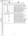
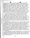
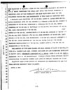
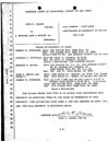
Manufacturer's Guide and Technical Data
Picture made with a Naessens Microscope
Back To Top Of Page

Click on thumbnail for larger image.
Back To Top Of Page